Beyond hypertrophy: Four-chamber myocardial involvement in ATTR cardiac amyloidosis diagnosed by multimodality imaging
DOI:
https://doi.org/10.21542/gcsp.2026.31Abstract
Background: Cardiac amyloidosis results from the deposition of amyloid fibrils in the myocardial extracellular matrix, leading to restrictive cardiomyopathy and heart failure. Diagnosis has traditionally been challenging owing to non-specific clinical features and reliance on endomyocardial biopsy. Advances in cardiovascular imaging have, however, improved early detection.
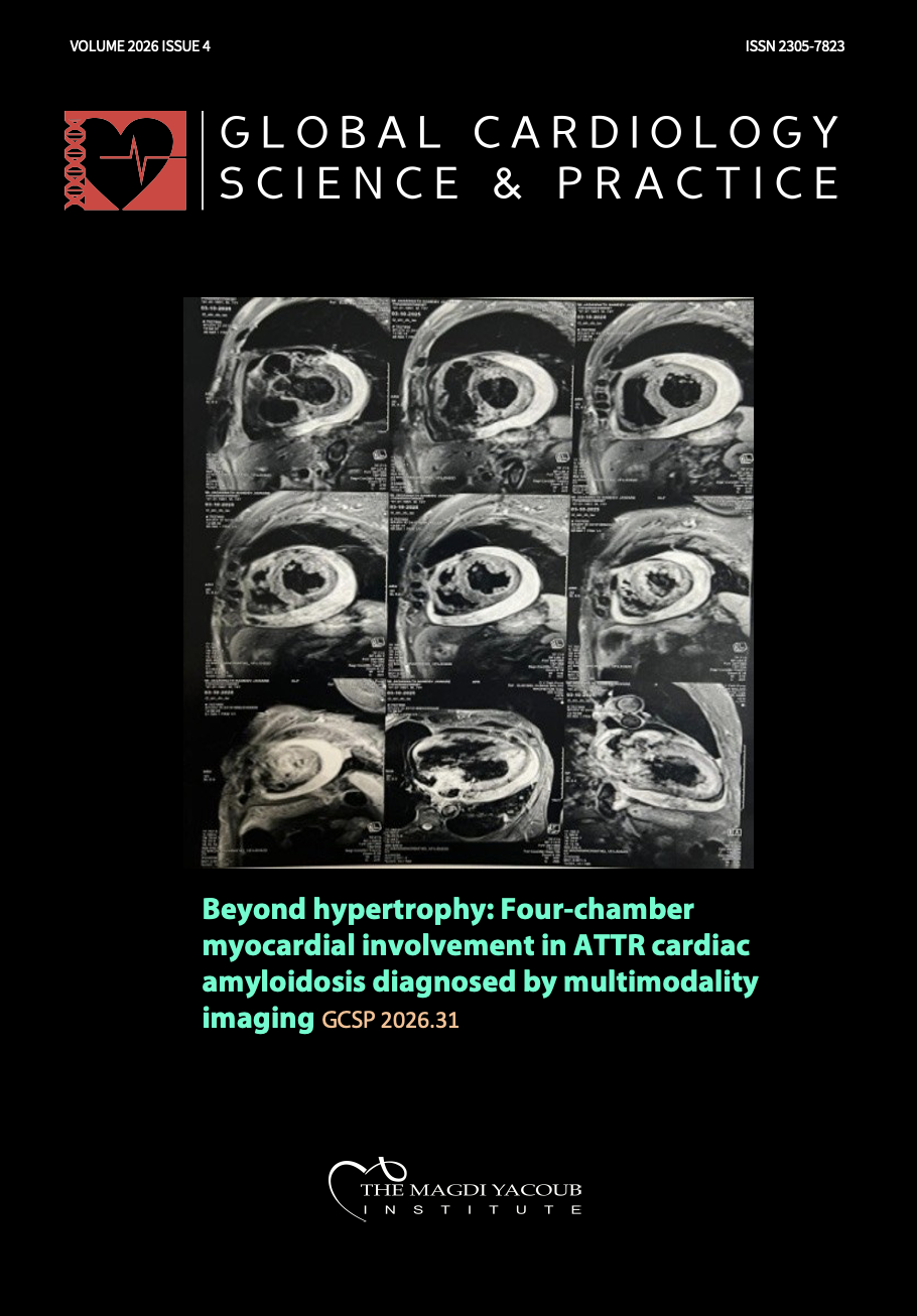
Case summary: We present a 74-year-old man with progressive dyspnea on exertion and bilateral pedal edema of three months' duration. Cardiac amyloidosis was suspected on the basis of discordance between electrocardiographic and echocardiographic findings, and was confirmed using global longitudinal strain (GLS), cardiac magnetic resonance imaging (CMR), and bone scintigraphy.
Conclusion: This case underscores the importance of recognizing cardiac amyloidosis as an underdiagnosed cause of heart failure. Modern imaging modalities facilitate non-invasive, early diagnosis, often obviating the need for biopsy. Early identification is crucial, as emerging therapies offer the potential to modify disease progression and improve outcomes in a condition once considered incurable.
Downloads
Published
Issue
Section
License
Copyright (c) 2026 Sravanth Gandavarapu

This work is licensed under a Creative Commons Attribution 4.0 International License.
This is an open access article distributed under the terms of the Creative Commons Attribution license CC BY 4.0, which permits unrestricted use, distribution and reproduction in any medium, provided the original work is properly cited.